A high ESR may indicate inflammation or infection; it is not a diagnosis on its own.
Educational guide only — not medical advice. Always review results with a qualified clinician.
6 min read
··Last updated
NNoryaAI
High ESR: what your blood test result means
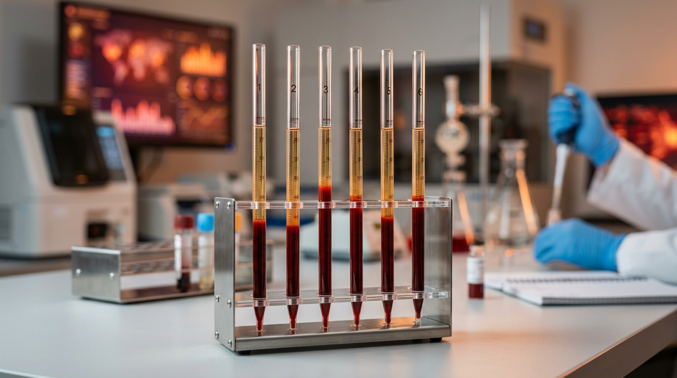
The ESR (Erythrocyte Sedimentation Rate) is one of the oldest and simplest blood tests still in routine use. It measures how quickly red blood cells (erythrocytes) settle to the bottom of a vertical tube over one hour. A faster settling rate indicates that inflammation is likely present in the body—though it does not pinpoint where or why.
Despite the availability of more specific inflammation markers such as C-reactive protein (CRP), the ESR remains valuable because it reflects chronic, low-grade inflammatory processes that CRP may not capture as well. It is widely used to help diagnose and monitor conditions like temporal arteritis, polymyalgia rheumatica, rheumatoid arthritis, and systemic lupus erythematosus.
This guide explains what the ESR measures, how to interpret your result, common causes of elevation, and when to seek medical attention. It is for educational purposes only and does not replace a doctor's advice.
What is ESR (Erythrocyte Sedimentation Rate)?
The ESR is a non-specific marker of inflammation. When inflammation is present in the body, the liver increases production of certain proteins—particularly fibrinogen and immunoglobulins—that cause red blood cells to stick together and form stacks called rouleaux. These rouleaux are heavier than individual red blood cells and therefore settle faster in the test tube, producing a higher ESR reading.
The test was first described in the early 1900s and standardized by Westergren in 1921. The Westergren method remains the reference standard: anticoagulated blood is drawn into a 200 mm vertical tube, and the distance the red cells have fallen from the top after exactly 60 minutes is recorded in millimeters per hour (mm/hr).
Because the ESR depends on changes in plasma proteins rather than detecting a specific pathogen or cytokine, it is considered a non-specific test. An elevated ESR tells your doctor that something is causing inflammation but requires additional investigation to determine the cause. Conversely, a normal ESR does not rule out disease entirely, as some inflammatory conditions may not elevate it significantly.
How does the ESR test work?
The physics behind ESR are straightforward. Red blood cells normally repel each other because they carry a net negative charge on their surface (the zeta potential). This negative charge keeps cells dispersed in plasma, causing them to settle slowly.
When acute-phase proteins such as fibrinogen increase during inflammation, they reduce the zeta potential between red cells, allowing cells to aggregate into rouleaux formations. These multi-cell stacks have a greater mass-to-surface-area ratio and therefore sink faster through the plasma column. The rate of sedimentation is proportional to the concentration of these aggregation-promoting proteins.
Several factors besides inflammation can alter the ESR. Anemia increases ESR because fewer red cells create less resistance to sedimentation. Polycythemia (excess red cells) has the opposite effect, slowing sedimentation. Abnormally shaped red cells (sickle cells, spherocytes) do not form rouleaux well and tend to produce falsely low ESR values. The test is also affected by temperature, tube tilt, and time delay before processing—which is why standardized conditions are important for reliable results.
Normal ESR ranges
Group
Normal ESR (mm/hr)
Men < 50 years
0 – 15
Men ≥ 50 years
0 – 20
Women < 50 years
0 – 20
Women ≥ 50 years
0 – 30
A commonly used age-adjusted formula is: upper limit of normal = age/2 for men and (age+10)/2 for women. This acknowledges that ESR naturally rises with age due to increased fibrinogen levels and other plasma protein changes.
An ESR above 100 mm/hr is considered markedly elevated and strongly suggests serious underlying disease, such as infection, malignancy, or autoimmune disease. Values between 40–100 are moderately elevated and warrant investigation. Mildly elevated values (20–40) are common and may reflect minor inflammation, aging, or obesity.
Causes of high ESR
An elevated ESR has a broad differential diagnosis. The most important categories include:
Chronic infections tend to cause higher ESR elevations than acute viral infections.
Autoimmune and inflammatory diseases:
Temporal (giant cell) arteritis and polymyalgia rheumatica – ESR is a key diagnostic criterion; values often exceed 50–100 mm/hr.
Rheumatoid arthritis – ESR correlates with disease activity.
Systemic lupus erythematosus (SLE) – ESR rises during flares, though CRP may remain normal in SLE (a useful distinguishing feature).
Inflammatory bowel disease, vasculitis, and sarcoidosis.
Malignancies: Multiple myeloma, lymphoma, and metastatic cancers can cause markedly elevated ESR. In myeloma, the excess immunoglobulins strongly promote rouleaux formation.
Other causes: Anemia, pregnancy (ESR normally rises in the second and third trimester), end-stage renal disease, heart failure, obesity, and advanced age.
ESR vs. CRP: which inflammation marker is better?
Both ESR and CRP are markers of inflammation, but they behave differently and provide complementary information:
Feature
ESR
CRP
Response speed
Slow (days to change)
Fast (rises within 6–8 hours)
Normalization
Slow (weeks)
Rapid (days)
Affected by anemia
Yes (falsely elevated)
No
Best for
Chronic inflammation monitoring
Acute infection detection
In systemic lupus erythematosus, the ESR often rises while CRP remains normal—this discordance helps distinguish SLE flares from infections. For more on CRP, see our CRP guide.
In practice, many clinicians order both tests together to get a more complete picture of a patient's inflammatory status.
False elevations and limitations of ESR
Because ESR depends on red blood cell behavior and plasma protein composition, several non-inflammatory conditions can falsely elevate the result:
Anemia – lower hematocrit means less resistance to sedimentation, producing a falsely high ESR even without inflammation.
Aging – ESR rises 1–2 mm/hr per decade of age due to increasing fibrinogen.
Female sex – women tend to have higher ESR than men.
Pregnancy – physiological increase in fibrinogen and plasma volume.
Kidney failure – uremia alters plasma proteins.
Hypergammaglobulinemia – high immunoglobulins (as in myeloma or liver disease) strongly promote rouleaux formation.
Conditions that can falsely lower ESR include polycythemia, sickle cell disease, spherocytosis, extreme leukocytosis, and certain protein abnormalities. Clinicians must interpret ESR in the context of the patient's overall clinical picture.
The role of ESR in disease monitoring
While ESR has limited value as a screening tool in asymptomatic patients, it is extremely useful for monitoring disease activity over time in conditions where it is known to correlate with clinical status:
Temporal arteritis / polymyalgia rheumatica – ESR is followed serially during treatment with corticosteroids. A rising ESR may indicate relapse.
Rheumatoid arthritis – ESR is part of composite disease activity scores (DAS28). Declining ESR suggests treatment is working.
Infections – ESR tracks the response to antibiotic therapy in chronic infections like osteomyelitis or endocarditis.
Hodgkin lymphoma – ESR is an independent prognostic factor; high ESR at diagnosis is associated with worse outcomes.
Because ESR changes slowly, it is best suited for tracking chronic conditions rather than acute changes. CRP is preferred when rapid response to treatment needs to be assessed. The combination of both markers provides the most informative longitudinal tracking of inflammatory disease.
When to see a doctor
Consult your doctor about an elevated ESR in the following situations:
Your ESR is above the age-adjusted normal range and you have unexplained symptoms such as fatigue, weight loss, fever, or joint pain.
Your ESR is above 100 mm/hr—this strongly suggests serious underlying disease and requires prompt evaluation.
You have been diagnosed with an autoimmune or inflammatory condition and your ESR is rising despite treatment.
You have new-onset severe headache, jaw claudication, or visual changes in patients over 50—these may indicate temporal arteritis, a medical emergency.
You have persistent unexplained anemia with high ESR, which may suggest multiple myeloma or other malignancy.
A mildly elevated ESR in an otherwise healthy person may not require immediate action but should be noted and rechecked if symptoms develop. Context is everything when interpreting this test.
How Norya helps you understand your ESR results
Understanding your ESR result alongside other blood markers can be challenging. Norya simplifies this: upload your blood test results and receive a clear, structured health summary within minutes. Norya evaluates your ESR in the context of CRP, complete blood count, and other inflammation markers to help you see the full picture.
The report highlights abnormal values, explains their significance in plain language, and helps you prepare the right questions for your doctor. Start your free analysis with Norya.
Disclaimer
This guide is for informational purposes only and does not replace medical advice or diagnosis. Always discuss your results with a healthcare professional. Start analysis with Norya
Trust & review
How this guide should be used
This article is educational and should be reviewed alongside our medical review, methodology, and transparency pages. Use it to prepare for a clinician conversation, not as a diagnosis.