Platelets are essential for blood clotting; a high or low count alone is not a diagnosis.
Educational guide only — not medical advice. Always review results with a qualified clinician.
7 min read
··Last updated
NNoryaAI
PLT (Platelet) test: What your results mean
Your PLT (platelet count) is one of the key values reported on a complete blood count (CBC). Platelets — also called thrombocytes — are tiny, disc-shaped cell fragments produced by megakaryocytes in the bone marrow. Their primary job is to stop bleeding by forming clots at the site of vascular injury.
A platelet count that is higher than normal (thrombocytosis) or lower than normal (thrombocytopenia) can signal a wide range of medical conditions — from infections and iron deficiency to autoimmune diseases and bone marrow disorders. Understanding your platelet count in context is essential for identifying potential health concerns early.
This comprehensive guide explains what platelets are, how they work, what normal values look like, causes of abnormal counts, the relationship between PLT and MPV, symptoms to watch for, and when to consult a doctor. This article is for educational purposes only and does not replace professional medical advice.
What are platelets (thrombocytes)?
Platelets (thrombocytes) are small, anucleate cell fragments that circulate in the bloodstream. They are produced in the bone marrow by giant precursor cells called megakaryocytes. A single megakaryocyte can shed thousands of platelets into the circulation. Each platelet is only about 2–3 micrometres in diameter — far smaller than red or white blood cells — yet they play an outsized role in maintaining vascular integrity.
Platelets have a lifespan of approximately 7–10 days. Once they age, they are removed primarily by the spleen, which also serves as a reservoir holding roughly one-third of the body's total platelet pool. This is why conditions that enlarge the spleen (splenomegaly) can sequester platelets and reduce the circulating count.
Beyond hemostasis, platelets contribute to innate immunity by releasing antimicrobial peptides and interacting with leukocytes. They also secrete growth factors (e.g., PDGF, TGF-β) that promote wound healing, tissue repair, and angiogenesis. Platelet-rich plasma (PRP) therapy leverages these regenerative properties in orthopedic and dermatologic medicine.
How platelets work in blood clotting
When a blood vessel is damaged, the underlying collagen is exposed, and platelets immediately adhere to the injured surface. This initial contact is mediated by von Willebrand factor (vWF), a glycoprotein that acts as molecular glue between platelet receptors (GP Ib/IX/V) and collagen. Upon adhesion, platelets become activated: they change from smooth discs to spiny spheres with pseudopodia that interlock with one another.
Activated platelets release the contents of their dense granules — ADP, thromboxane A2 (TXA2), and serotonin — which recruit additional platelets to the site. Simultaneously, the GP IIb/IIIa receptors on the platelet surface undergo a conformational change that allows them to bind fibrinogen, cross-linking adjacent platelets. This process is called aggregation and produces the primary hemostatic plug, which can stop bleeding from small vessels within minutes.
The coagulation cascade then reinforces this initial plug. Tissue factor activates a chain of clotting factors that ultimately generates thrombin, which converts soluble fibrinogen into insoluble fibrin strands. The fibrin mesh stabilizes the platelet plug, forming the definitive secondary hemostatic clot. Any defect in platelet number or function — whether too few, too many, or dysfunctional — can tip the balance toward excessive bleeding or pathologic thrombosis.
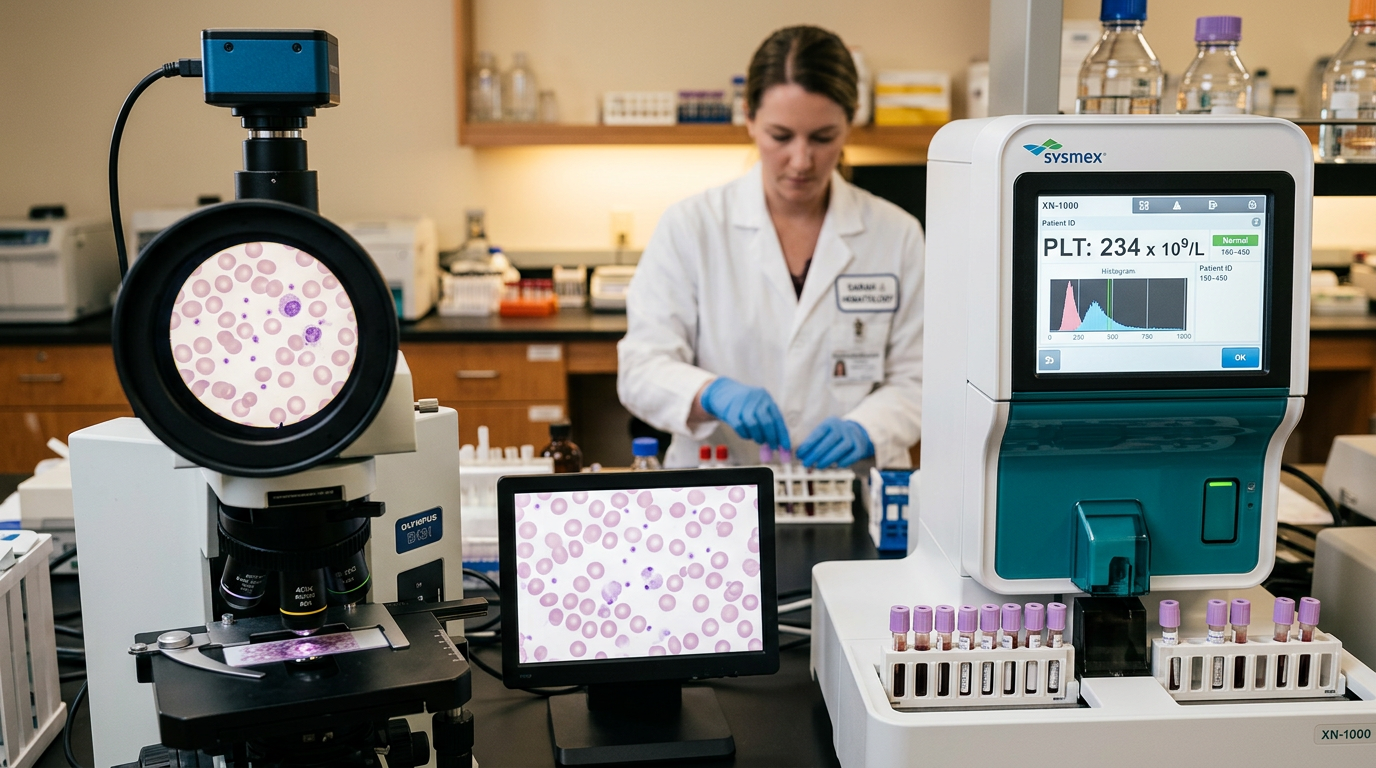
Normal platelet (PLT) ranges
Platelet count is measured as part of the complete blood count (CBC) and is reported in thousands per microlitre (×10³/μL) or cells per microlitre:
Parameter
Normal range
Note
PLT (Adults)
150,000–400,000/μL
Same for both sexes
PLT (Neonates)
150,000–450,000/μL
Slightly wider range in the first weeks
MPV (Mean Platelet Volume)
7.5–12.0 fL
Indicates platelet size
Platelet counts can fluctuate physiologically throughout the day. Transient increases occur after vigorous exercise, stress, and epinephrine release. A mild decrease may be observed during menstruation. High altitude living can also elevate counts. Some individuals may exhibit pseudothrombocytopenia — a falsely low count caused by EDTA-dependent platelet clumping in the collection tube. If suspected, a repeat sample drawn into a citrate tube usually resolves the discrepancy.
Reference intervals can vary slightly between laboratories; always compare your result to the reference range printed on your specific lab report.
Causes of high platelets (thrombocytosis)
Thrombocytosis is defined as a platelet count exceeding 400,000/μL. It is classified into two major categories:
1. Reactive (secondary) thrombocytosis accounts for the vast majority of cases and occurs as a response to an underlying condition:
Infections — Acute bacterial, viral, or fungal infections stimulate megakaryopoiesis through cytokines such as IL-6 and thrombopoietin.
Iron deficiency anemia — Iron deficiency stimulates thrombopoiesis through cross-reactivity of thrombopoietin receptors with erythropoietin pathways.
Surgery and trauma — Postoperative acute-phase response causes transient thrombocytosis, typically peaking 1–2 weeks after the event.
Splenectomy — Removal of the spleen releases sequestered platelets and removes the primary site of platelet clearance, often causing persistent elevation.
Malignancy — Some cancers (lung, ovarian, GI) can cause paraneoplastic thrombocytosis.
2. Primary (clonal) thrombocytosis arises from a stem-cell-level defect in the bone marrow. Myeloproliferative neoplasms — essential thrombocythemia (ET), polycythemia vera (PV), and chronic myeloid leukemia (CML) — fall into this category. Mutations in JAK2, CALR, or MPL genes are commonly identified. Primary thrombocytosis carries a heightened risk of both thrombotic and hemorrhagic complications.
Causes of low platelets (thrombocytopenia)
Thrombocytopenia is defined as a platelet count below 150,000/μL. It can result from three broad mechanisms:
1. Decreased production (bone marrow suppression):
Aplastic anemia — Bone marrow failure affecting all cell lineages.
Leukemia, lymphoma, and metastatic cancer — Malignant infiltration crowds out normal megakaryopoiesis.
Immune thrombocytopenic purpura (ITP) — Autoimmune antibodies target platelet surface glycoproteins, leading to accelerated splenic destruction. Acute ITP is common in children following viral illness; chronic ITP predominates in adults.
Thrombotic thrombocytopenic purpura (TTP) — ADAMTS13 deficiency causes microangiopathic hemolytic anemia and severe thrombocytopenia.
Heparin-induced thrombocytopenia (HIT) — An antibody-mediated paradoxical prothrombotic state triggered by heparin therapy.
Disseminated intravascular coagulation (DIC) — Widespread activation of clotting cascades consumes platelets and clotting factors simultaneously.
3. Splenic sequestration: When the spleen enlarges due to liver cirrhosis, portal hypertension, or infiltrative diseases (e.g., Gaucher disease), it traps a larger proportion of platelets, reducing the circulating count. Drug-induced thrombocytopenia (quinidine, valproic acid, sulfonamides, certain antibiotics, and chemotherapy agents) is also a frequently encountered cause that should not be overlooked.
MPV (Mean Platelet Volume) and its connection to PLT
MPV (Mean Platelet Volume) measures the average size of platelets in femtolitres (fL). The normal range is typically 7.5–12.0 fL. MPV provides insight into the rate of platelet production and the state of the bone marrow.
High MPV + low PLT: This pattern suggests that the bone marrow is compensating for peripheral platelet loss by releasing younger, larger platelets. It is characteristically seen in immune thrombocytopenic purpura (ITP) and other consumptive causes. Young platelets contain more granules and are hemostatically more active than older, smaller ones.
Low MPV + low PLT: This combination may indicate that the bone marrow itself is failing to produce adequate platelets — as seen in aplastic anemia, post-chemotherapy suppression, and myelodysplastic syndromes. When the marrow cannot mount a compensatory response, both count and size remain depressed.
Elevated MPV has also been linked to increased cardiovascular risk. Larger platelets are more prone to aggregation and have been identified as an independent risk factor for myocardial infarction, stroke, and venous thromboembolism. For more details on MPV, see our MPV article.
Symptoms of high and low platelet counts
Thrombocytopenia (low PLT) symptoms:
Petechiae — Pinpoint, flat red-purple spots on the skin that do not blanch with pressure.
Purpura — Larger areas of subcutaneous bleeding, appearing as purple patches.
Easy bruising (ecchymosis) — Bruises that appear from minor or no trauma.
Gum bleeding — Excessive bleeding during brushing or flossing.
Nosebleeds (epistaxis) — Frequent and hard-to-stop nasal bleeding.
Heavy menstrual bleeding (menorrhagia) — Abnormally prolonged or heavy periods.
GI or urinary bleeding — In severe thrombocytopenia, melena, hematuria, or hematemesis may occur.
Thrombocytosis (high PLT) symptoms: Reactive thrombocytosis is usually asymptomatic; the symptoms of the underlying condition dominate. However, in primary thrombocytosis (e.g., essential thrombocythemia), the following may occur:
Erythromelalgia — Burning pain, warmth, and redness in the hands and feet.
Headache and visual disturbances — Due to microvascular occlusion.
Thrombosis — Deep vein thrombosis (DVT), pulmonary embolism, stroke, or myocardial infarction.
Paradoxical bleeding — At very high counts (>1,000,000/μL), acquired von Willebrand disease can develop, causing bleeding rather than clotting.
Spontaneous bleeding risk increases significantly when the platelet count drops below 50,000/μL. Below 10,000–20,000/μL, life-threatening internal hemorrhage — including intracranial bleeding — becomes a serious concern requiring urgent medical attention.
When to see a doctor
Consult a healthcare professional promptly if any of the following apply:
Your platelet count is below 150,000/μL or above 400,000/μL.
You notice unexplained petechiae, purpura, or bruises on your skin.
You experience frequent or prolonged nosebleeds or gum bleeding.
Post-surgical or post-dental bleeding takes an unusually long time to stop.
You have unexplained burning, pain, or redness in your hands or feet (erythromelalgia).
Your platelet count shows a significant upward or downward trend compared to previous tests.
Your doctor may order additional investigations, including a peripheral blood smear, bone marrow biopsy, immature platelet fraction (IPF), thrombopoietin levels, or genetic testing (JAK2, CALR, MPL mutations) to determine the underlying cause. Early diagnosis is particularly important in hematologic malignancies, where timely treatment can significantly improve outcomes.
If your platelet count is critically low (<20,000/μL) and you have signs of active bleeding, this is a medical emergency — seek immediate care at the nearest emergency department.
How Norya helps you understand your results
By uploading your blood test to Norya, you can receive a structured, easy-to-understand health summary of your PLT, MPV, and other hematologic parameters within minutes. Norya compares your values against reference ranges, highlights potential deviations, and presents your results in a holistic perspective.
This summary is an ideal tool for preparing for your doctor's appointment: you can clearly see which values need attention, how related parameters connect, and what your overall picture looks like. Norya does not diagnose — but it empowers you to understand your results and have a more productive conversation with your physician.
Disclaimer
This guide is for informational purposes only and does not replace medical advice or diagnosis. Always discuss your results with a healthcare professional. Start analysis with Norya
Trust & review
How this guide should be used
This article is educational and should be reviewed alongside our medical review, methodology, and transparency pages. Use it to prepare for a clinician conversation, not as a diagnosis.